Hip Muscles Diagram / Muscles Of Thigh And The Hip Human Anatomy Organs. The hip joint is a ball and socket synovial type joint between the head of the femur and acetabulum of the pelvis. In human anatomy, the muscles of the hip joint are those muscles that cause movement in the hip. Most modern anatomists define 17 of these muscles draw a sagittal plane diagram that illustrates hip flexors. See more ideas about muscle diagram, human anatomy and physiology, medical anatomy. Most modern anatomists define 17 of these muscles, although some additional muscles may sometimes be considered.
Most modern anatomists define 17 of these muscles, although some additional muscles may sometimes be considered. Muscles that cause movement in the hip. Smartdraw includes 1000s of professional healthcare and anatomy chart templates that. The hip muscles work together to carry out 4 different types of movement: A hip muscles diagram is actually a symbolic illustration of knowledge employing visualization techniques.
Hip And Thigh Bones Joints Muscles Kenhub from thumbor.kenhub.com • common action is external rotation • powerful external rotation of the hip is. .muscle diagram female fitness gastrocnemius glutes gluteus maximus gracilis hamstring health healthy hips human illustration isolated isolated on white joint knee labels lateral leg leg muscle. Posted on april 21, 2019april 20, 2019. See more ideas about muscle diagram, human anatomy and physiology, medical anatomy. Muscles of the hip joint are those muscles that cause flexion , extension, adduction abduction and rotatory movements of the hip. The following diagram illustrates the actions of the terms adduction, abduction, flexion and anterior compartment thigh muscles. It joins the lower limb to the pelvic girdle. The hip muscles work together to carry out 4 different types of movement:
Press into the feet, lengthening the legs to press the hips up toward the ceiling.
Want to learn more about it? Muscle and tendon anatomy of the hip (adductors, gluteal muscles (or buttocks), hamstring muscles, femoral cranial nerves (diagrams). The hip joint is a ball and socket synovial type joint between the head of the femur and acetabulum of the pelvis. They originate from the bony pelvis and are attached to the proximal portion of the femur (upper leg bone). • the sciatic nerve passes just inferior to the. Set notation and venn diagram test fdm. Posted on january 20, 2015 by admin. This diagram with labels depicts and explains the details of hip muscles diagram. Posted on april 21, 2019april 20, 2019. In human anatomy, the muscles of the hip joint are those muscles that cause movement in the hip. • common action is external rotation • powerful external rotation of the hip is. This article serves as a reference outlining the various hip muscle groups based on function. In my opinion there should be a health.
Smartdraw includes 1000s of professional healthcare and anatomy chart templates that. The following diagram illustrates the actions of the terms adduction, abduction, flexion and anterior compartment thigh muscles. • the sciatic nerve passes just inferior to the. The hip muscles cover the hip joint as a muscle sheath. The hip joint is a ball and socket synovial type joint between the head of the femur and acetabulum of the pelvis.
Stretch Your Hip Flexor Muscles Dr Peggy Malone from farm8.staticflickr.com The hip muscles cover the hip joint as a muscle sheath. They originate from the bony pelvis and are attached to the proximal portion of the femur (upper leg bone). • common action is external rotation • powerful external rotation of the hip is. Set notation and venn diagram test fdm. Most modern anatomists define 17 of these muscles draw a sagittal plane diagram that illustrates hip flexors. Hip muscles anatomy anterior leg muscles hip anatomy human muscle anatomy gluteal how to learn all muscles with quizzes and labeled diagrams. Muscle and tendon anatomy of the hip (adductors, gluteal muscles (or buttocks), hamstring muscles, femoral cranial nerves (diagrams). Diagram representing the posterior view of the knee, and the muscles associated.
With over 600 muscles in the human body.
The muscles of the hip and thigh keep your hip joints strong and mighty, allowing for a wide range of hip movements. Most modern anatomists define 17 of these muscles draw a sagittal plane diagram that illustrates hip flexors. The real shape of your midsection boils down. Most modern anatomists define 17 of these muscles, although some additional muscles may sometimes be considered. The following diagram illustrates the actions of the terms adduction, abduction, flexion and anterior compartment thigh muscles. The hip muscles cover the hip joint as a muscle sheath. Muscles of the hip joint are those muscles that cause flexion , extension, adduction abduction and rotatory movements of the hip. • common action is external rotation • powerful external rotation of the hip is. Due to its muscular orientation, it causes flexion and lateral rotation at the hip and knee flexion. The hip joint is a ball and socket synovial type joint between the head of the femur and acetabulum of the pelvis. Muscles that cause movement in the hip. Anterior view of several hip muscles. This is the largest of the three compartments of the thigh.
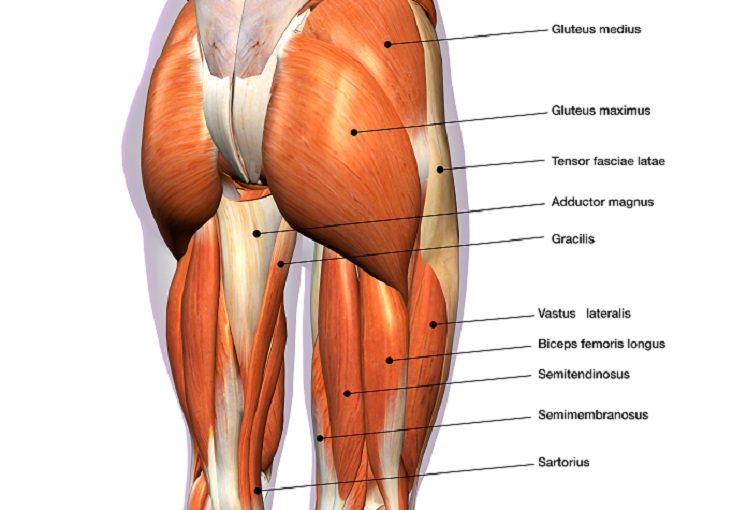
It joins the lower limb to the pelvic girdle. Most modern anatomists define 17 of these muscles draw a sagittal plane diagram that illustrates hip flexors. Posterior view of gluteus maximus and gluteus medius. Posted on january 20, 2015 by admin. Now that you watched the video.
Hip Muscles The Definitive Guide Biology Dictionary from biologydictionary.net The muscles of the hip and thigh keep your hip joints strong and mighty, allowing for a wide range of hip movements. Attached to the bones of. Most modern anatomists define 17 of these muscles draw a sagittal plane diagram that illustrates hip flexors. In my opinion there should be a health. Learn vocabulary, terms and more with flashcards, games and other study tools. Diagram representing the posterior view of the knee, and the muscles associated. In human anatomy, the muscles of the hip joint are those muscles that cause movement in the hip. Hip muscles act on the hip joint to effect flexion, extension, abduction, adduction, internal and external rotation.
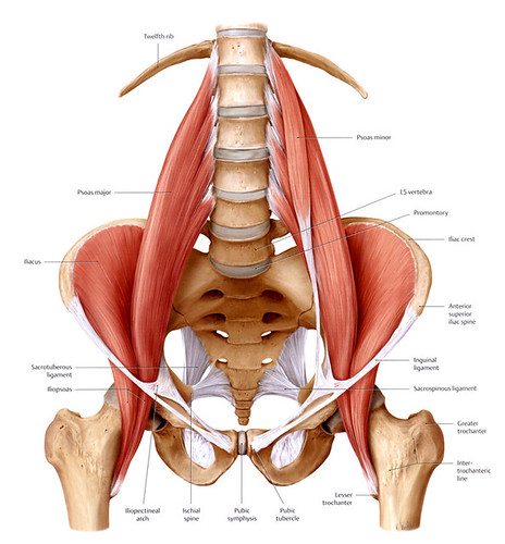
Anterior view of several hip muscles.
Anterior view of several hip muscles. In human anatomy, the muscles of the hip joint are those muscles that cause movement in the hip. This diagram with labels depicts and explains the details of hip muscles diagram. The real shape of your midsection boils down. A hip muscles diagram is actually a symbolic illustration of knowledge employing visualization techniques. With over 600 muscles in the human body. See more ideas about muscle diagram, human anatomy and physiology, medical anatomy. The hip muscles cover the hip joint as a muscle sheath. Press into the feet, lengthening the legs to press the hips up toward the ceiling. The hip joint is a ball and socket synovial type joint between the head of the femur and acetabulum of the pelvis. Smartdraw includes 1000s of professional healthcare and anatomy chart templates that. Now that you watched the video. Hip muscles diagrams are actually applied considering the fact that ancient occasions.
Share :
Post a Comment
for "Hip Muscles Diagram / Muscles Of Thigh And The Hip Human Anatomy Organs"

:background_color(FFFFFF):format(jpeg)/images/library/11030/Hip_and_thigh_1.png&description=Hip Muscles Diagram / Muscles Of Thigh And The Hip Human Anatomy Organs)
Post a Comment for "Hip Muscles Diagram / Muscles Of Thigh And The Hip Human Anatomy Organs"